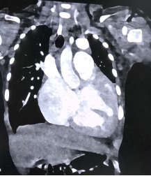
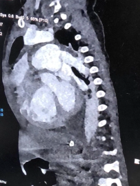
Total anomalous pulmonary venous connection (TAPVC) is a rare cyanotic congenital heart defect where all pulmonary veins drain into systemic veins rather than the left atrium, impairing oxygenation and risking heart failure or pulmonary hypertension, thus necessitating surgery; when coexisting with Factor VII deficiency—an autosomal recessive disorder impairing the extrinsic coagulation pathway—surgical bleeding risk increases, requiring multidisciplinary planning. We report a 7-year-old girl from Jammu and Kashmir, India, presenting with cyanosis during crying, where echocardiography and cardiac catheterization confirmed coronary sinus-type TAPVC and a 14 mm ostium secundum atrial septal defect (OS-ASD) that partially relieved right atrial pressure; she had congenital Factor VII deficiency (14.8% activity preoperatively), demanding vigilant coagulopathy monitoring. In 2024, she developed a brain abscess and left forearm osteomyelitis, treated from February 18 to April 1, with burrhole drainage for the abscess revealing the Factor VII deficiency via coagulopathy workup. Readmitted on October 28, 2025, she underwent surgery on November 26, 2025, involving unroofing of the coronary sinus to redirect pulmonary veins to the left atrium and OS-ASD patch closure via midline sternotomy under cardiopulmonary bypass; prothrombin time and fibrinogen levels remained stable intra- and postoperatively, eliminating the need for recombinant Factor VIIa (rFVIIa), with postoperative Factor VII activity at 13%. She tolerated weaning from inotropes and ventilation without complications, had sutures removed, and was discharged on December 7, 2025, on enalapril for afterload reduction, sildenafil for pulmonary vasodilation, furosemide for diuresis, and amiodarone for rhythm control. This case illustrates successful management of moderate Factor VII deficiency in complex pediatric cardiac surgery without hemostatic prophylaxis, given minimal bleeding and rigorous monitoring, providing insights for resource-limited settings and emphasizing cardiothoracic-haematology collaboration for optimal outcomes.
| Published in | International Journal of Cardiovascular and Thoracic Surgery (Volume 12, Issue 2) |
| DOI | 10.11648/j.ijcts.20261202.17 |
| Page(s) | 63-69 |
| Creative Commons |
This is an Open Access article, distributed under the terms of the Creative Commons Attribution 4.0 International License (http://creativecommons.org/licenses/by/4.0/), which permits unrestricted use, distribution and reproduction in any medium or format, provided the original work is properly cited. |
| Copyright |
Copyright © The Author(s), 2026. Published by Science Publishing Group |
Total Anomalous Pulmonary Venous Connection, Factor VII Deficiency, Pediatric Cardiac Surgery, Cyanosis, Coronary Sinus Tapvc, No Replacement Therapy
TAPVC | Total Anomalous Pulmonary Venous Connection |
OS-ASD | Ostium Secundum Atrial Septal Defect |
TAPVR | Total Anomalous Pulmonary Venous Return |
RV | Right Ventricle |
CPB | Cardiopulmonary Bypass |
PVO | Pulmonary Venous Obstruction |
rFVIIa | Recombinant Activated FVII |
fVII | Factor VII |
CTA | Computed Tomography Angiography |
INR | International Normalised Ratio |
PT | Prothrombin Time |
MRI | Magnetic Resonance Imaging |
TTE | Transthoracic Echocardiography |
TEE | Transesophageal Echocardiography |
CHD | Congenital Heart Disease |
CS | Coronary Sinus |
POD | Post Operative Day |
STAT | Society of Thoracic Surgeons-European Association for Cardio-Thoracic Surgery |
TEG | Thromboelastography |
| [1] | Hammon JW, Bender HW, Graham TP, Cotton RB, Hazinski MF, Christian K, et al., Surgical management of total anomalous pulmonary venous connection: Thirty-four years' experience. J Thorac Cardiovasc Surg. 1992; 104(6): 1505-11. |
| [2] | Harada T, Nakano T, Oda S, Kado H., Surgical results of total anomalous pulmonary venous connection: 25 years' experience. Interact Cardiovasc Thorac Surg. 2019; 28(3): 421-6. |
| [3] | Karamlou T, Gurofsky R, Al Abbas G, Williams WG, Freedom RM, Blackstone EH, et al., Surgery for total anomalous pulmonary venous connection: Outcomes and predictors of survival. J Thorac Cardiovasc Surg. 2007; 133(4): 1046-52. |
| [4] | Shi G, Zhu Z, Chen J, Ou Y, Hong H, Nie Z, et al., Contemporary Outcomes of Surgical Repair of Total Anomalous Pulmonary Venous Connection in Neonates and Infants. Ann Thorac Surg. 2015; 100(2): 659-65. |
| [5] | Li J, Wu Z, Zhang J, Wang Y, He B., Comparison of the Efficacy and Safety of Sutureless Technique and Conventional Surgery for Total Anomalous Pulmonary Venous Connection: A Systematic Review and Meta-Analysis. Front Cardiovasc Med. 2024; 11: 41522630. |
| [6] | Nahle AA, Hamdar H, Soqia J, Diab M, Ataya J, Al-Dairy A., Factors associated with early postoperative mortality after total anomalous pulmonary venous connection repair. Medicine (Baltimore). 2024; 103(21): e38788. |
| [7] | Wen C, Shi G, Zhang Q, Zhu F, Zhang H, Zhu Z, et al., Review of surgical experience in 61 patients with mixed total anomalous pulmonary venous connection. Eur J Cardiothorac Surg. 2022; 61(6): 1299-1305. |
| [8] | Sakamoto T, Nagashima M, Umezu K, Houki R, Ikarashi J, Katagiri J, et al., Long-term outcomes of total correction for isolated total anomalous pulmonary venous connection. Interact Cardiovasc Thorac Surg. 2018; 27(1): 20-6. |
| [9] | Seale AN, Uemura H, Webber SA, Partridge J, Roughton M, Ho SY, et al., Outcome of postoperative pulmonary venous obstruction after repair of total anomalous pulmonary venous connection. J Thorac Cardiovasc Surg. 2012; 145(5): 1255-62. |
| [10] | Milovanovic V, Mimic B, Vulicevic I, Divac I, Parezanovic V, Ilisic T, et al., Outcomes of surgery for total anomalous pulmonary venous drainage: A single-center experience. Srp Arh Celok Lek. 2014; 142(3-4): 164-9. |
| [11] | Yousuf S, Faisal F, Amna A, et al., Perioperative management of severe factor VII deficiency: A case report. PMC - NIH. 2024; PMC12497768. |
| [12] | Nandi S, Karki S., Congenital factor VII deficiency: Multidisciplinary approach is the key to success. PMC - NIH. 2017; PMC5372415. |
| [13] | Ozkan S, Senol S, et al., Perioperative Management of Patients with Rare Congenital Factor Deficiency. EJMI. 2023; 7(1): 82-87. |
| [14] | El Ansari T, Mamad H, Benkirane S, Masrar A., Perioperative Challenges in a Patient With Moderate Congenital Factor VII Deficiency: A Case Report. Cureus. 2024; 16(11): e43164. |
| [15] | Chon HN, Cho JH, Park YS., A Retrospective Review at a Single Hemophilia Treatment Center: Coagulation Factor VII Deficiency. Clin Pediatr Hematol Oncol. 2020; 27(2): 113-119. |
| [16] | Omer M, Soneji N., Severe Congenital Factor VII Deficiency with Normal Perioperative Hemostasis: A Case Report. Am J Case Rep. 2021; 22: e930245. |
| [17] | Khan MA, Ali S., Anesthetic Management of a Pediatric Patient with Factor VII Deficiency undergoing Orchidopexy. J Neonatal Surg. 2019; 8: 86-11. |
| [18] | Li Y, Zhang L., A new perspective on perioperative coagulation management in Factor VII deficiency. Medicine (Baltimore). 2018; 97(44): e13023. |
| [19] | Subramaniam S, Kumar S., Cardiac type of total anomalous pulmonary venous connection: Anatomy and Repair. PMC. 2013; PMC3603772. |
| [20] | Yan L, Zhou Y, Li D, Li L, Tang H., Case report: Thoughts on two cases of total anomalous pulmonary venous connection. Front Cardiovasc Med. 2023; 10: 1075168. |
| [21] | White BR, Ho VT, Guleserian KJ., Cardiac-type total anomalous pulmonary venous return is not benign. J Thorac Cardiovasc Surg. 2022; 164(2): e11-13. |
| [22] | Luo X, Wang H, Wang J., Novel Repair for Obstructed Total Anomalous Pulmonary Venous Connection. Ann Thorac Surg. 2003; 75(4): 1322-1324. |
| [23] | Kabbani SS, Jureidini S, Canter C., TOTAL ANOMALOUS PULMONARY VENOUS CONNECTION TO CORONARY SINUS. J Am Coll Cardiol. 2025; 85(4): 421-430. |
| [24] | Zhang M, Wang L, Zhao H., Prognostic factors in pediatrics TAPVC: a 10-year retrospective study. Sci Rep. 2025; 15: 94619. |
| [25] | Lee J, Kim Y, Park S., Total anomalous pulmonary venous return with mixed drainage and a rare connection to the azygos vein. Eur Heart J Case Rep. 2022; 6(11): ytac415. |
| [26] | Ahmed S, Gupta R., Total Anomalous Pulmonary Venous Connection with Rare Direct Connection to the Right Atrium. PMC - NIH. 2024; PMC11419768. |
| [27] | Ozcan V, Demir S., Totally Anomalous Pulmonary Venous Connection Outcomes from A Single Center: 10 Years Experience. Kosuyolu Heart J. 2023; 26(1): 18-24. |
| [28] | Sharma R, Jain N., Coagulation Profile in Neonates with Congenital Heart Disease. PMC. 2024; PMC10890703. |
| [29] | Warren O, et al., Recombinant Activated Factor VII in Cardiac Surgery: A Systematic Review. Ann Thorac Surg. 2007; 83(2): 707-14. |
| [30] | Raksamani K, et al., Use of Recombinant Activated Factor VII for Controlling Refractory Postoperative Bleeding in Pediatric Cardiac Surgery. J Cardiothorac Vasc Anesth. 2011; 25(6): 1001-1006. |
| [31] | Omer M, Soneji N., Severe Congenital Factor VII Deficiency with Normal Perioperative Hemostasis. Am J Case Rep. 2021; 22: e936657. |
| [32] | Taylor S, Miller P., Prothrombin Complex Concentrate vs Factor VII for Refractory Bleeding After Cardiac Surgery. Ann Thorac Surg Short Rep. 2023; 1(1): 220-6. |
| [33] | Guzzetta NA., Current and future trends in coagulation management for congenital heart surgery. J Thorac Cardiovasc Surg. 2017; 154(3): 1038-1042. |
| [34] | Gill R, et al., Safety and Efficacy of Recombinant Activated Factor VII in Cardiac Surgery. Circulation. 2009; 120(1): 21-27. |
| [35] | Knio ZO, et al., Undiagnosed Factor VII Deficiency in Cardiac Surgery Complicated by Bleeding: A Case Report. A&A Pract. 2023; 17(9): e01713. |
| [36] | Sugimoto T, et al., Open-heart surgery in an infant with heterozygous factor VII deficiency. Interact Cardiovasc Thorac Surg. 2010; 10(6): 1037-9. |
| [37] | Mariani G, et al., A retrospective analysis of 157 surgical procedures performed in 56 patients with Type I congenital factor VII deficiency. J Thromb Haemost. 2022; 20(6): 698-703. |
| [38] | Omer M., Severe Congenital Factor VII Deficiency with Normal Perioperative Hemostasis and Blood Loss. PMC. 2021; PMC8366573. |
| [39] | European Medical Journal., Management of Patients with Factor VII Deficiency in Surgery: A Comprehensive Review. EMJ. 2022; 44(2): 222-224. |
| [40] | Diplaris K, et al., Treatment of refractory bleeding after cardiac operations with low-dose recombinant activated factor VII. Eur J Cardiothorac Surg. 2008; 33(1): 64-9. |
| [41] | Wang Z, Ma K, Li S., Long-Term Outcomes of Surgical Repair of Supracardiac Total Anomalous Pulmonary Venous Connection. Pediatr Cardiol. 2024; 45(5): 389-182. |
| [42] | Lund-Olesen S, et al., Nationwide registry study of long term survival and comorbidities in total anomalous pulmonary venous connection. Sci Rep. 2025; 15: 15769. |
| [43] | Zhu Z, et al., Outcomes of total anomalous pulmonary venous drainage repair in a mid-volume center. JTCVS Open. 2022; 11: 360-6. |
| [44] | Seder WB, et al., Long-Term Transplant-Free Survival After Repair of Total Anomalous Pulmonary Venous Connection. PMC. 2017; PMC5729081. |
| [45] | Bhardwaj V, et al., Analysis of Risk Factors Associated with Early Mortality in Total Anomalous Pulmonary Venous Connection (TAPVC) Repairs. J Card Crit Care. 2024; 8: 54. |
| [46] | Noda T, et al., Arrhythmic Burden of Adult Survivors With Repaired Total Anomalous Pulmonary Venous Return. JACC Adv. 2022; 1(1): 277-281. |
| [47] | Shi G, et al., Contemporary Outcomes of Surgical Repair of Total Anomalous Pulmonary Venous Connection in 202 Patients. Ann Thorac Surg. 2015; 99(6): 2134-40. |
| [48] | Korbmacher B, et al., Long-term results after repair of total anomalous pulmonary venous connection. Thorac Cardiovasc Surg. 2001; 49(2): 101-6. |
| [49] | Seale AN, et al., Total Anomalous Pulmonary Venous Connection: Anatomy, Clinical Features, and Management. Circulation. 2010; 122(25): 2718-26. |
APA Style
Satpathy, A. S., Pavan, M., Pramanik, B., Mishra, A. K., Bansal, D. (2026). Successful Surgical Correction of Coronary Sinus Total Anomalous Pulmonary Venous Connection Without rFVIIa Intervention in Congenital Factor VII Deficiency. International Journal of Cardiovascular and Thoracic Surgery, 12(2), 63-69. https://doi.org/10.11648/j.ijcts.20261202.17
ACS Style
Satpathy, A. S.; Pavan, M.; Pramanik, B.; Mishra, A. K.; Bansal, D. Successful Surgical Correction of Coronary Sinus Total Anomalous Pulmonary Venous Connection Without rFVIIa Intervention in Congenital Factor VII Deficiency. Int. J. Cardiovasc. Thorac. Surg. 2026, 12(2), 63-69. doi: 10.11648/j.ijcts.20261202.17
AMA Style
Satpathy AS, Pavan M, Pramanik B, Mishra AK, Bansal D. Successful Surgical Correction of Coronary Sinus Total Anomalous Pulmonary Venous Connection Without rFVIIa Intervention in Congenital Factor VII Deficiency. Int J Cardiovasc Thorac Surg. 2026;12(2):63-69. doi: 10.11648/j.ijcts.20261202.17
@article{10.11648/j.ijcts.20261202.17,
  author = {Abhishek Soham Satpathy and Mallikarjuna Pavan and Biswarup Pramanik and Anand Kumar Mishra and Deepak Bansal},
  title = {Successful Surgical Correction of Coronary Sinus Total Anomalous Pulmonary Venous Connection Without rFVIIa Intervention in Congenital Factor VII Deficiency},
  journal = {International Journal of Cardiovascular and Thoracic Surgery},
  volume = {12},
  number = {2},
  pages = {63-69},
  doi = {10.11648/j.ijcts.20261202.17},
  url = {https://doi.org/10.11648/j.ijcts.20261202.17},
  eprint = {https://article.sciencepublishinggroup.com/pdf/10.11648.j.ijcts.20261202.17},
  abstract = {Total anomalous pulmonary venous connection (TAPVC) is a rare cyanotic congenital heart defect where all pulmonary veins drain into systemic veins rather than the left atrium, impairing oxygenation and risking heart failure or pulmonary hypertension, thus necessitating surgery; when coexisting with Factor VII deficiency—an autosomal recessive disorder impairing the extrinsic coagulation pathway—surgical bleeding risk increases, requiring multidisciplinary planning. We report a 7-year-old girl from Jammu and Kashmir, India, presenting with cyanosis during crying, where echocardiography and cardiac catheterization confirmed coronary sinus-type TAPVC and a 14 mm ostium secundum atrial septal defect (OS-ASD) that partially relieved right atrial pressure; she had congenital Factor VII deficiency (14.8% activity preoperatively), demanding vigilant coagulopathy monitoring. In 2024, she developed a brain abscess and left forearm osteomyelitis, treated from February 18 to April 1, with burrhole drainage for the abscess revealing the Factor VII deficiency via coagulopathy workup. Readmitted on October 28, 2025, she underwent surgery on November 26, 2025, involving unroofing of the coronary sinus to redirect pulmonary veins to the left atrium and OS-ASD patch closure via midline sternotomy under cardiopulmonary bypass; prothrombin time and fibrinogen levels remained stable intra- and postoperatively, eliminating the need for recombinant Factor VIIa (rFVIIa), with postoperative Factor VII activity at 13%. She tolerated weaning from inotropes and ventilation without complications, had sutures removed, and was discharged on December 7, 2025, on enalapril for afterload reduction, sildenafil for pulmonary vasodilation, furosemide for diuresis, and amiodarone for rhythm control. This case illustrates successful management of moderate Factor VII deficiency in complex pediatric cardiac surgery without hemostatic prophylaxis, given minimal bleeding and rigorous monitoring, providing insights for resource-limited settings and emphasizing cardiothoracic-haematology collaboration for optimal outcomes.},
 year = {2026}
}
TY  - JOUR
T1  - Successful Surgical Correction of Coronary Sinus Total Anomalous Pulmonary Venous Connection Without rFVIIa Intervention in Congenital Factor VII Deficiency
AU  - Abhishek Soham Satpathy
AU  - Mallikarjuna Pavan
AU  - Biswarup Pramanik
AU  - Anand Kumar Mishra
AU  - Deepak Bansal
Y1  - 2026/03/27
PY  - 2026
N1  - https://doi.org/10.11648/j.ijcts.20261202.17
DO  - 10.11648/j.ijcts.20261202.17
T2  - International Journal of Cardiovascular and Thoracic Surgery
JF  - International Journal of Cardiovascular and Thoracic Surgery
JO  - International Journal of Cardiovascular and Thoracic Surgery
SP  - 63
EP  - 69
PB  - Science Publishing Group
SN  - 2575-4882
UR  - https://doi.org/10.11648/j.ijcts.20261202.17
AB  - Total anomalous pulmonary venous connection (TAPVC) is a rare cyanotic congenital heart defect where all pulmonary veins drain into systemic veins rather than the left atrium, impairing oxygenation and risking heart failure or pulmonary hypertension, thus necessitating surgery; when coexisting with Factor VII deficiency—an autosomal recessive disorder impairing the extrinsic coagulation pathway—surgical bleeding risk increases, requiring multidisciplinary planning. We report a 7-year-old girl from Jammu and Kashmir, India, presenting with cyanosis during crying, where echocardiography and cardiac catheterization confirmed coronary sinus-type TAPVC and a 14 mm ostium secundum atrial septal defect (OS-ASD) that partially relieved right atrial pressure; she had congenital Factor VII deficiency (14.8% activity preoperatively), demanding vigilant coagulopathy monitoring. In 2024, she developed a brain abscess and left forearm osteomyelitis, treated from February 18 to April 1, with burrhole drainage for the abscess revealing the Factor VII deficiency via coagulopathy workup. Readmitted on October 28, 2025, she underwent surgery on November 26, 2025, involving unroofing of the coronary sinus to redirect pulmonary veins to the left atrium and OS-ASD patch closure via midline sternotomy under cardiopulmonary bypass; prothrombin time and fibrinogen levels remained stable intra- and postoperatively, eliminating the need for recombinant Factor VIIa (rFVIIa), with postoperative Factor VII activity at 13%. She tolerated weaning from inotropes and ventilation without complications, had sutures removed, and was discharged on December 7, 2025, on enalapril for afterload reduction, sildenafil for pulmonary vasodilation, furosemide for diuresis, and amiodarone for rhythm control. This case illustrates successful management of moderate Factor VII deficiency in complex pediatric cardiac surgery without hemostatic prophylaxis, given minimal bleeding and rigorous monitoring, providing insights for resource-limited settings and emphasizing cardiothoracic-haematology collaboration for optimal outcomes.
VL  - 12
IS  - 2
ER  -
Cardiac Thoracic Vascular Surgery Department, Post Graduate Institute of Medical Education and Research, Chandigarh, India
Cardiac Thoracic Vascular Surgery Department, Post Graduate Institute of Medical Education and Research, Chandigarh, India
Cardiac Thoracic Vascular Surgery Department, Post Graduate Institute of Medical Education and Research, Chandigarh, India
Cardiac Thoracic Vascular Surgery Department, Post Graduate Institute of Medical Education and Research, Chandigarh, India
Clinical Hematology, Post Graduate Institute of Medical Education and Research, Chandigarh, India